Lung
Cancer Screening Using Low-Dose CT
What is cancer screening?
Screening is the use of tests or examinations to detect a disease in people
without symptoms of that disease. For example, colonoscopy is used for
colon cancer screening. Screening for cancers is important because
it can help the doctor discover cancer early and treat it successfully.
Because lung cancer usually spreads beyond the lungs before causing
any symptoms, an effective screening program for early detection of lung
cancer has the potential to save many lives.
Studies of the use of chest X-rays for lung cancer screening in the 1970s
and 1980s concluded that chest x-rays could not find many lung cancers
early enough to improve a patients chance for a cure. For this reason,
lung cancer screening was not a routine practice for the general public or
even for people at increased risk, such as smokers.
In the past decade or so, low-dose CT
(computerized tomography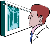 ) scans of the
chest have been successful in detecting
early lung cancers in smokers and former
smokers. Because this test is useful among
people at high risk for lung cancer, the
Worker Health Protection Program (WHPP)
offers annual low dose CT scans to
program participants aged 50 to 85 who
have a history of smoking and occupational exposure to known lung
cancer risk factors, such as asbestos, radiation and beryllium. WHPP
participants aged 50 to 85 are also eligible if the medical screening chest
X-ray results show scarring of the lung related to asbestos or silica, or the
participant has chronic beryllium disease. ) scans of the
chest have been successful in detecting
early lung cancers in smokers and former
smokers. Because this test is useful among
people at high risk for lung cancer, the
Worker Health Protection Program (WHPP)
offers annual low dose CT scans to
program participants aged 50 to 85 who
have a history of smoking and occupational exposure to known lung
cancer risk factors, such as asbestos, radiation and beryllium. WHPP
participants aged 50 to 85 are also eligible if the medical screening chest
X-ray results show scarring of the lung related to asbestos or silica, or the
participant has chronic beryllium disease.
Why is screening for Lung Cancer
in a high-risk population so Important?
Lung cancer is the leading cause of cancer death for both men and
women. About 130,000 people in the United States will die of lung cancer
each year. Without screening, over 50 percent of lung cancers are found
at a late stage and the overall five-year survival rate for lung cancer is
currently 20 percent, meaning only 20 of every 100 people survive at least
five years. By contrast, if lung cancer is found early and treated by
surgery, before it has spread to lymph nodes or other organs, the fiveyear survival rate increases dramatically to 70 percent or higher. This
means that at least 70 out of 100 of these patients are likely to survive for
at least five years.
Studies have shown that low-dose CT screening detects many lung tumors at
early stages. For example, a 1999 Early Lung Cancer Action Program (ELCAP)
study of 1,000 smokers and former smokers, low-dose chest CT found 27
tumors while conventional chest X-rays detected only 7 cancers; 23 of the 27
CT-detected tumors (85 percent) were in the early stages. The X-rays only
found 4 early tumors.
After the ELCAP study, several large, randomized clinical studies in the U.S.
and Europe confirmed the effectiveness of low-dose CT. One of the largest was
the 2011 National Cancer Institute (NCI)s National Lung Screening Trial
(NLST) that showed a 20 percent reduction in deaths from lung cancer among
current and former smokers who underwent annual low-dose chest CT
screenings for 3 to 5 years, compared to a similar group who underwent chest
X-ray screenings.
What is spiral, low-dose CT?
Low-dose spiral CT is a simple procedure in which a special imaging machine
rotates rapidly around the body taking over 100 pictures in sequence. This
information is processed by a computer to produce a cross-section of a specific
area. The low-dose CT scan uses less radiation than a standard CT and yet is
sensitive enough to detect abnormalities that are too small to be seen on a
conventional set of chest X-rays.
How is the low-dose CT procedure done?
Throughout the low-dose CT scanning procedure, the patient lies very still on a
table. The patient passes through the x-ray machine, which is shaped like a
doughnut with a large hole. The machine rotates around the patient and a
computer creates images from the scan that can be reconstructed into a 3-
dimensional model of the lungs. When the picture is taken, you will be asked to
hold your breath for approximately 10 seconds. The amount of radiation (an
estimated average of 1.2 mSv or 120 mrem for most people) is significantly less
than that absorbed during a diagnostic CT scan of the chest (an estimated
average of 7 mSv or 700 mrem). As further comparison, the estimated average
annual exposure from natural sources in soil and air is 3.1 mSv or 310 mrem
per year.
What will happen if the CT scan shows an abnormality in my lung?
If an abnormality is detected on the CT scan, it may or may not be cancer. If
there is suspicion of a lung cancer, you will be notified within 10 working days
of the screening. At that time, we will advise you to see your personal physician,
who may recommend diagnostic testing. We do not pay for any such diagnostic
testing or any treatment that may follow. Those expenses are normally covered
by health insurance including Medicaid and Medicare. Sometimes the doctor
can tell from the CT scan that the lung nodule is not cancerous. In this case, or
if the CT scan shows no nodule at all, you will be notified within four weeks of
the screening, and no follow-up will be needed.
|